


For people with amputation who have prosthetic limbs, one of the greatest challenges is controlling the prosthesis so that it moves the same way a natural limb would. Most prosthetic limbs are controlled using electromyography, a way of recording electrical activity from the muscles, but this approach provides only limited control of the prosthesis.
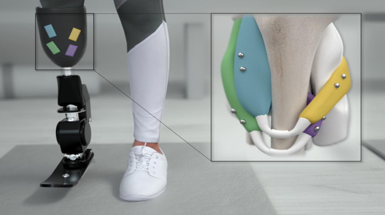
U.S. Researchers have now developed an alternative approach that they believe could offer much more precise control of prosthetic limbs. After inserting small magnetic beads into muscle tissue within the amputated residuum, they can precisely measure the length of a muscle as it contracts, and this feedback can be relayed to a bionic prosthesis within milliseconds.
In a new study appearing today in Science Robotics, the researchers tested their new strategy, called magnetomicrometry (MM), and showed that it can provide fast and accurate muscle measurements in animals. They hope to test the approach in people with amputation within the next few years.
“Hopefully, MM will replace electromyography as the dominant way to link the peripheral nervous system to bionic limbs. MM has a high signal quality, has a low regulatory hurdle and cost, and is minimally invasive.”
Senior Researcher
The new strategy is based on the idea that if sensors could measure what muscles are doing, those measurements would offer more precise control of a prosthesis. To achieve, that, the researchers decided to insert pairs of magnets into muscles. By measuring how the magnets move relative to one another, the researchers can calculate how much the muscles are contracting and the speed of contraction.
Using an array of magnetic sensors placed on the outside of the legs, the researchers found that they were able to determine the position of the magnets with a precision of 37 microns (about the width of a human hair), as they moved the turkeys’ ankle joints. These measurements could be obtained within three milliseconds.
For control of a prosthetic limb, these measurements could be fed into a computer model that predicts where the patient’s phantom limb would be in space, based on the contractions of the remaining muscle. This strategy would direct the prosthetic device to move the way that the patient wants it to, matching the mental picture that they have of their limb position.
With magnetomicrometry, researchers are directly measuring the length and speed of the muscle. Through mathematical modelling of the entire limb, they can compute target positions and speeds of the prosthetic joints to be controlled, and then a simple robotic controller can control those joints.
Within the next few years, the researchers hope to do a small study on human patients who have amputations below the knee. They envision that the sensors used to control the prosthetic limbs could be placed on clothing, attached to the surface of the skin, or affixed to the outside of a prosthesis.
MM could also be used to improve the muscle control achieved with a technique called functional electrical stimulation, which is now used to help restore mobility in people with spinal cord injuries. Another possible use for this kind of magnetic control would be to guide robotic exoskeletons, which can be attached to an ankle or another joint to help people who have suffered a stroke or developed other kinds of muscle weakness.
U.S. Researchers have been developing a variety of technologies to help people with disabilities and diseases. As reported by OpenGov Asia, A new robotic neck brace from U.S. researchers may help doctors analyse the impact of cancer treatments on the neck mobility of patients and guide their recovery.
The new brace was made using 3D-printed materials and inexpensive sensors. The easy-to-wear device was based on the head and neck movements of 10 healthy individuals. This is the first study of this kind where a wearable robotic neck brace has been designed to characterize the full head and neck range of motion.
In the future, the researchers hope to investigate larger groups of patients and use the neck brace to follow patients through physical therapy to develop evidence-based protocols for rehabilitation. They also would like to develop similar braces for other surgical sites, such as the forearm, ankle, or knee.


