


Scientists from Nanyang Technological University have developed luminescent imaging probes that could allow for early detection of acute kidney failure.
This health condition is mostly common among patients who are critically ill and need intensive care. Current measures for diagnosing kidney failure are unable to detect the pre-morbid changes in the early stage which indicate acute renal failure.
The sensitivity of this probe allows for faster tracking of changes in the biological process which are triggered by the onset of the condition.
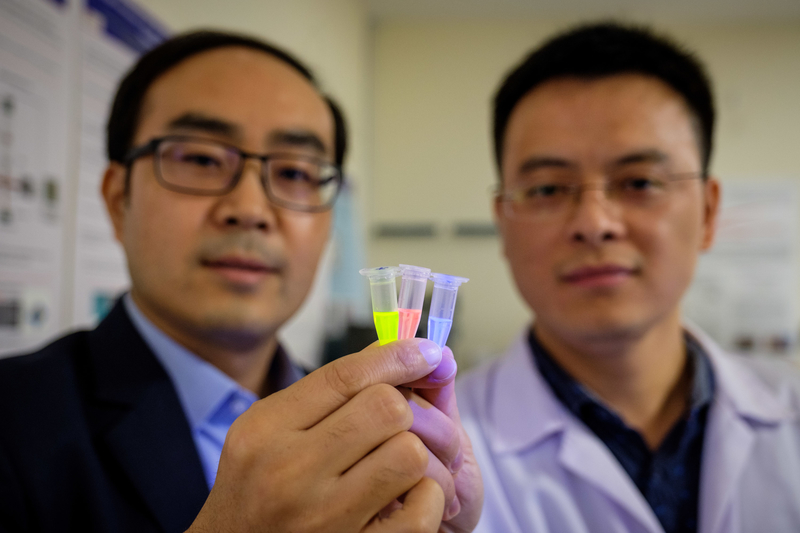
These probes have been developed by Associate Professor Pu Kanyi and his team from NTU.
NTU Associate Professor Pu Kanyi said, “For patients who are critically ill, like those in the intensive care unit, every minute is precious in reversing a condition like acute kidney failure, which can cause a patient’s health to deteriorate rapidly”.
How these probes work
The imbalance of reactive oxygen species (ROS), which are chemically unstable molecules in the body that act as early-stage indicators, leads to damage in the body’s fatty tissues, DNA, and proteins. This can result in cell deaths that can no longer be healed by an injured kidney.
The team created probes into three components:
- Part that reacts with the identified ROS
- Luminescence signalling part that ensures the probe lights up
- Part that ensures the probe passes through the kidneys instead of accumulating in the liver
These renal probes are injected into the bloodstream and after a period, they ‘light up’ when they detect molecular changes caused by the onset of acute kidney failure. In the study done on the mice, the probes lit up 12 hours after injecting the mice with the cancer drug, cisplatin, which is destructive to the kidneys.
Acute kidney failure commonly occurs in a few hours or days. The probes tested on the mice took 1.5 days earlier than the current molecular imaging process to detect.
The team of scientists have also found the probes to have a high renal clearance where more than 97 percent of the probes injected into the mice flow through the kidneys and are being excreted through urine.
This allows for a non-intrusive way of detecting kidney failure. The probes can be re-developed as test strips and directly added to urine samples. After a few hours of incubation, the probes will light up when exposed to UV light in the presence of biomarkers.


