


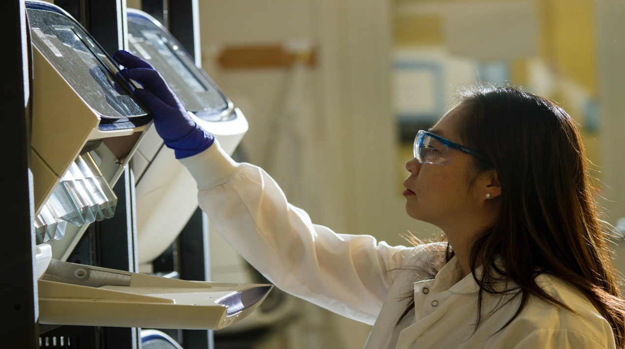
An AI-powered analytic tool to predict the effectiveness of a complementary biomarker for immune checkpoint inhibition in non-small-cell lung cancer (NSCLC) has been developed by Professor Tony MOK Shu Kam from The Chinese University of Hong Kong’s (CUHK) Faculty of Medicine (CU Medicine) in collaboration with South Korea’s Seoul National University College of Medicine, Sungkyunkwan University School of Medicine, Ajou University School of Medicine and an AI start-up.
The tool conducts a spatial analysis of the distribution of tumour infiltrating lymphocytes biomarkers in whole slide images which can predict treatment outcomes with immune checkpoint inhibitors, the current first-line therapy for advanced NSCLC.
Study results showed that the AI-powered analysis of tumour infiltrating lymphocytes correlates with tumour response and progression-free survival, meaning it may help optimise treatment selection in clinical practice. These findings were recently published in the Journal of Clinical Oncology.
Immune checkpoint inhibitors are standard therapy for advanced NSCLC with PD-L1 expression
Lung cancer is one of the most common cancers in the world, accounting for 1.8 million deaths every year. According to the Hong Kong cancer registry, lung cancer is the leading cause of cancer deaths and the most common cancer in the city, with more than 5,000 new cases every year. NSCLC accounts for above 80% of all lung cancers.
Immune checkpoint inhibitors are a standard first-line therapy for advanced NSCLC with PD-L1 expression, known as anti-PD-1 checkpoint inhibitors. When protein PD-1 on immune cell T-cell binds with ligand PD-L1 from cancer cells, it prevents the immune system from killing cancer cells. The inhibitors serve the purpose of preventing the binding from taking place and allowing the immune system to do its work and kill cancer cells.
Professor Tony Mok, Li Shu Fan Professor of Clinical Oncology and Chairman of the Department of Clinical Oncology at CU Medicine stated that the outcome of anti-PD-1 checkpoint inhibitors may vary depending on the patient’s tumour microenvironment but there is currently no standard biomarker addressing this.
He noted that tumour infiltrating lymphocytes, in theory, are the main activator of antitumour immunity which could be a promising biomarker for predicting treatment outcomes with immune checkpoint inhibitors. But the current quantification method is labour-intensive and relies on spatial distribution in whole-slide images, limiting utility and objectivity.
Immune phenotypes correlating with tumour response to immune checkpoint inhibitors
The AI-powered spatial tumour infiltrating lymphocytes analyser developed by the collaborative research team is capable of segmentation and quantification of multiple histologic components from whole-slide images, including cancer epithelium, cancer stroma and tumour infiltrating lymphocytes.
Then, through deep learning-based AI model training, three immune phenotypes: inflamed, immune-excluded and immune-desert are generated. (Please refer to table 1 in the appendix for the definition of the three immune phenotypes)
Findings showed that the inflamed immune phenotype correlates with enrichment in local immune cytolytic activity, a higher response rate, and prolonged progression-free survival compared with patients with the other two phenotypes.
According to Professor Mok, this is the first study on AI-powered automated tumour infiltrating lymphocytes analysis in advanced NSCLC. In this study, the team has proved that the AI-powered spatial tumour infiltrating lymphocytes analyser is capable of predicting clinical outcomes of immune checkpoint inhibitors in patients with NSCLC. This may serve as a complementary biomarker to tumour proportion score, the current PD-L1 measurement.


