


A new revolutionary 3D coloured medical
scanner has scanned its first human. The scanner was invented by a tandem of
father and son scientists from the University of Canterbury and University of Otago
in New Zealand.
According to the report
made by the University of Canterbury, the MARS spectral x-ray scanner produces
images with significantly improved diagnostic information.
It measures the x-ray spectrum to produce
colour images instead of black-and-white ones, and shows different components
of body parts such as fat, water, calcium, and disease markers.
The scanner provides far greater detail of
the body’s chemical components, improving on the existing medical imaging,
which can change the diagnosis and treatments of diseases such as cancer and
heart disease.
Professor Phil Butler and his son,
Professor Anthony Butler are the scientists behind the MARS spectral x-ray
scanner. Professor Phil Butler is a physicist working at the University of
Canterbury while his son, Professor Anthony Butler is a radiologist and a
Professor at both the University of Otago and the University of Canterbury.
Professor Anthony Butler differentiated the
coloured images from the black-and-white ones saying that the x-ray spectral
information allows health professionals to measure the different components of
body parts such as fat, water, calcium, and disease markers, whereas
traditional black-and-white x-rays only allow measurement of the density and shape
of an object.
A technology used by the European
Organisation for Nuclear Research (CERN) was adapted by the Butlers as they turned
the ‘God particle’ into a medical scanner.
Professor Butler explained that it is the
Medpix3 technology of CERN which sets the machine apart diagnostically because
its small pixels and accurate energy resolution mean it can get images no other
imaging tool can.
Various research institutions around the
world already have small versions of the scanner that can house tissue samples.
The smaller version of the MARS scanner is
being used by researchers for the study of cancer, bone and joint health, and
vascular diseases that can cause heart attacks and strokes.
These studies have produced promising early
results suggesting that when spectral imaging is regularly used in clinics, it
will enable more accurate diagnosis and personalisation of treatment.
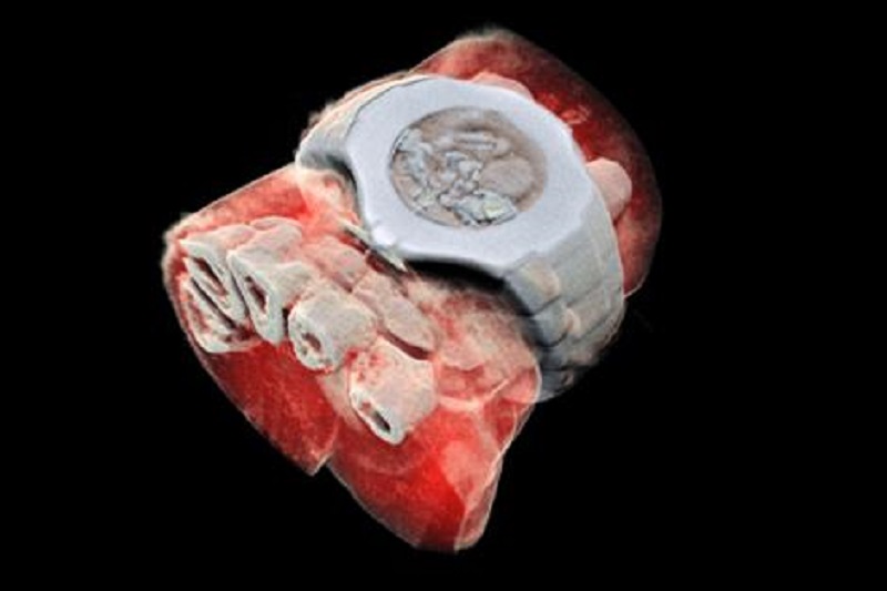
A larger form of the scanner had scanned
its first human. Professor Phil Butler, himself, was scanned. His ankle and
wrist were imaged.
More patients will get to try the scanner
in the coming months as the next step in development is an imminent clinical
trial where orthopaedic and rheumatology patients from Christchurch will be
scanned. The world-first clinical trial will allow the MARS team to compare the
images produced by their scanner with the technology currently used in New
Zealand hospitals.
Professor Anthony Butler explained that
after a decade in development, it is really exciting to have reached a point
where it is clear that the technology could be used for routine patient care.
He likened the new imaging scanner to a new
microscope wherein biomedical researchers can see different kinds of details
inside patients in a non-invasive manner.
Support has been given to the Butlers and
their growing team of scientists, during their decade-long development of the
machine, by the University of Canterbury and the University of Otago, the Ministry of Business,
Innovation and Employment, and GE Healthcare. In fact, MARS
Bioimaging Ltd (MBI) has commercialised the product.


