


In years past, NUS taught pathology
manually – using specimens. Medical students would inspect samples in
transparent containers or pots. Each student would await their turn to inspect
the pot, which gradually made its way around the lecture room.
By the time the last student at the back
had a chance to inspect the sample, the teacher had already moved on to another
topic.
Then, in a different session, the class
will then learn, from a separate tutor, how these diseases look on microscopic
slides.
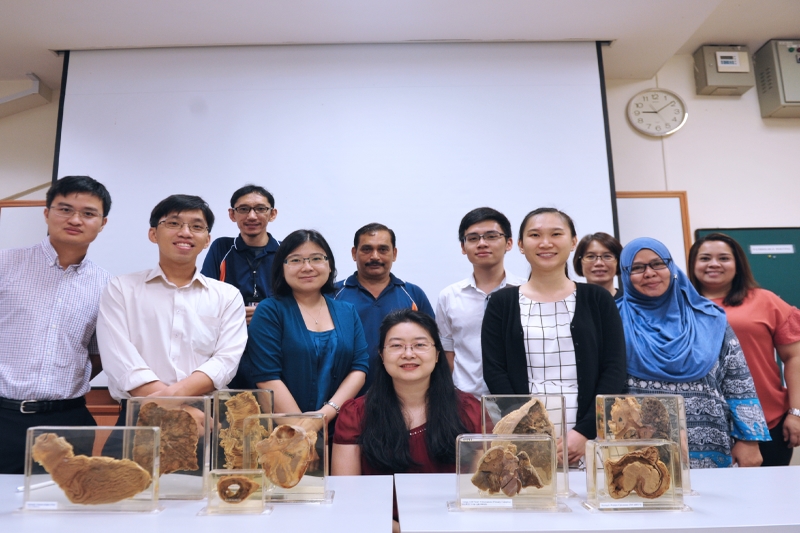
Today, however, NUS’s newsletter proudly reported
that the learning of pathology has been enhanced and aided through technology,
thanks to the efforts of Associate Professor Nga Min En and colleagues at the Department
of Pathology.
Painstakingly, one specimen at a time, the
team has rendered more than 700 specimens in digital format, making more than
250 specimens available online for medical students.
This labour
of love began more than six years ago and is still ongoing; scheduled to end
when the last and final specimen has been digitised,
said Assoc Prof Nga, who is a consulting pathologist at the National University
Hospital.
The benefits are greatly appreciated by
students, who no longer have to wait for their turn in class to view the
specimens, nor borrow them from the department to study for examinations.
The digitised
specimens of the diseased body parts can also be viewed alongside microscopic
slie images of the same disease, which helps students better understand the
morphology of diseases with more clarity.
Moreover, classes do not have to be split
into two groups too, since both the images of the disease and the specimens can
be viewed concurrently during lessons.
The digitisation
process

Photographing the samples and then converting
them to digitised images is done by a
team of non-academic staff at the Department of Pathology adept at IT and
photography.
The team also consists of students as well as NUHS Pathology
residents.
The latter help to check the teaching
materials for the online platform, provide ideas for improvement and make
value-added contributions such as annotations, adding links or cases.
The digitisation
process involves a carefully orchestrated photoshoot.
Staff position the specimens on a turntable
inside a lightbox, then photograph them
with an 18-megapixel camera at multiple
angles.
A total of 24 photos are taken of each
specimen.
Assoc Prof Nga then edits each image with
the Photoshop software, while another team member combines these 24 images into
a single file. The end result is a clear 360-degree view of the specimen in its
container.
This labour-intensive
process takes about 45 minutes per specimen.
Moving
online
Seeing the potential in going digital,
Assoc Prof Nga uploaded these materials online in 2015. Doing so enabled her
students to access them for their own learning.
While Assoc Prof Nga uses both physical and
virtual pots in her classes, she says that the digital pots offer greater flexibility
in terms of their use – students can study them any time they want to.
The updated web resource is called Pathweb and has two main sections.
In one section called the ‘Virtual
Pathology Museum', students can find both virtual microscopy slides and pots categorised into both general pathology and
systemic pathology, according to their curriculum.
Another section entitled ‘Pathology
Demystified’ features mind maps, live videos, talking slides and quizzes, all
hand-written, drawn or produced by Assoc Prof Nga.
These materials provide students with an
approach to studying pathology and allow
them to engage in self-directed learning.
Pathweb
and Assoc Prof Nga’s work have received acclaim and appreciation from students
and colleagues around the world.
The pathology department is home to more
than 5,000 dissected and preserved specimens of human organs and tissue.
While more than 700 of these pots have been
digitised since 2012, Assoc Prof Nga
hopes to digitise a further 1,000
specimens by the end of 2019, with the help of her team.


