


A team of researchers specialising in the field of imaging technology at the Department of Electrical and Electronic Engineering (EEE) at the University of Hong Kong (HKU) in collaboration with the University of California, Berkeley (UC Berkeley) has provided unprecedented moving images of how individual blood cells flow in the brain of a mouse that was awake.
The development provides neuroscientists with a tool to better understand the inner workings of the brain, especially how blood flow changes at the level of individual blood vessels and across the larger vessel network within the brain – an important cue to unravel how energy is distributed and regulated in healthy and diseased brains (e.g., Alzheimer’s disease, strokes).
The study, titled “Ultrafast two-photon fluorescence imaging of cerebral blood circulation in the mouse brain in vivo”, was published recently in the peer-reviewed Proceedings of the National Academy of Sciences.
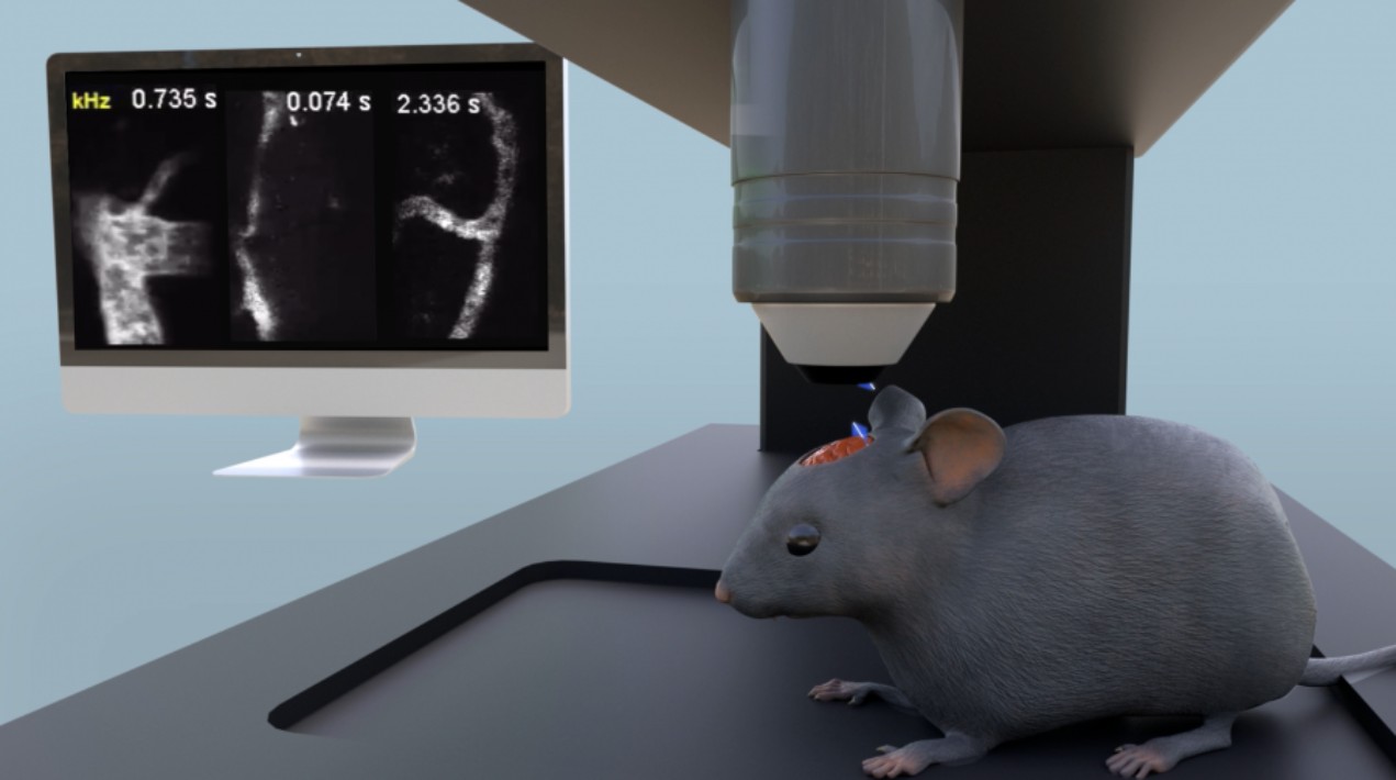
The team’s microscope has super high-speed two-photon fluorescence imaging that penetrates light more deeply into tissue than other gold-standard microscopy technologies to capture blood cells in fast motion. Most other technologies work with one-photon fluorescence and have a slow focus-scanning mechanism, which means they are limited to capturing low-flow speed regimens in anaesthetised animals that are not moving.
The two-photon imaging technology developed by the team, on the other hand, is able to capture faster moving flows – in this case, red blood cells – even when the animals are awake and able to move somewhat. The flow of red blood cells is an important cue of brain activity, which is fuelled by energy from the blood supply.
Professor Ji Na of UC Berkeley’s Department of Molecular & Cell Biology and Professor Kevin Tsia of HKU EEE who is also the Programme Director of the Biomedical Engineering Programme led the interdisciplinary research team, which includes engineers and neurobiologists.
Professor Tsia noted that a major limitation in other brain imaging technologies has been the resolution of images. The team’s technology can deal with these with very fast-moving images, at least 100 times faster than current state-of-the-art options, and capture them down to the individual blood cells. The cells can be counted, and their trajectories traced.
Images of the two approaches show stark differences. In the low-flow speed technology, the blood flow resembles a blur and smear. Images from the two-photon fluorescence technology are much more detailed and clearly show individual cells in fast motion.
The research team initially reported on their high-speed two-photon fluorescence technology in 2020 when they used it to successfully record the millisecond electrical signals of the neurons in an awake mouse. With this latest output, they have expanded the potential of their technology. The same technology has also been applied by Professor Tsia’s team in cancer screening through imaging cancer cells in blood.
The work on imaging individual blood cells in-vivo is an extension of that work and shows that the technology can be extended to other kinds of neuroscience research, especially cerebral haemodynamics, which is the dynamics of blood flow to the brain, Professor Tsia said.
The global medical imaging market is expected to grow from US$ 37.97 billion in 2021 to US$ 56.53 billion in 2028 at a CAGR of 5.8% in the forecast period, 2021-2028.


