


Laser light is used in the relatively new imaging technology known as photoacoustic microscopy (PAM) to cause ultrasonic vibrations in tissue. Like how ultrasound imaging functions, an image of the tissue’s architecture can then be produced using these ultrasonic vibrations and a computer that processes them.
Lihong Wang, the Bren Professor of Medical Engineering and Electrical Engineering at Caltech, has created PAM technologies recently that can visualise changing blood flow in the brain, identify specific cancer cells, and detect diseased tissue.
However, a drawback of PAM’s high resolution has been its shallow depth of field, which prevents it from focusing on more than one layer of tissue at once. This layer can only be as thin as one skin cell, or around 30 micrometres, with a resolution of one to two micrometres.
The device must refocus above or below the plane it is now observing to perceive objects above or below it. Consider someone donning reading glasses to do a crossword puzzle as a point of comparison.
The needle-shaped beam photoacoustic microscopy, or NB-PAM, that Wang and his research team created has a depth of field that is roughly 14 times larger than what was previously possible. As a result, NB-PAM can better photograph samples with uneven surfaces and produce 3-D pictures of samples without having to refocus.
According to Rui Cao, lead author and postdoctoral scholar research associate in medical engineering, certain applications, such as analysing tissue samples without requiring a microscope slide, need imaging of uneven surfaces at high spatial resolution. Hence, the new technology has addressed the trade-off between resolution and depth of field.
By adopting a longer, thinner, and more “needle-shaped” laser light beam than other PAM technologies, NB-PAM enhances the depth of field. By altering the optical properties of the beam, it is possible to avoid some of the problems that come with previous attempts to enhance the depth of field of PAM technology, such as slower operation or increased computing power needs.
A diffractive optical element (DOE) is a specialised tool used to produce this needle-shaped beam. A DOE appears to be a thin sheet of glass to the untrained eye, but it is a piece of fused silica with precise patterns carved on it.
These patterns alter the form of the imaging beam of light, causing it to be dragged out into a long, thin neck rather than focusing on a sharp point along the propagation axis. As a result, it can image objects more clearly at a wider range of depths.
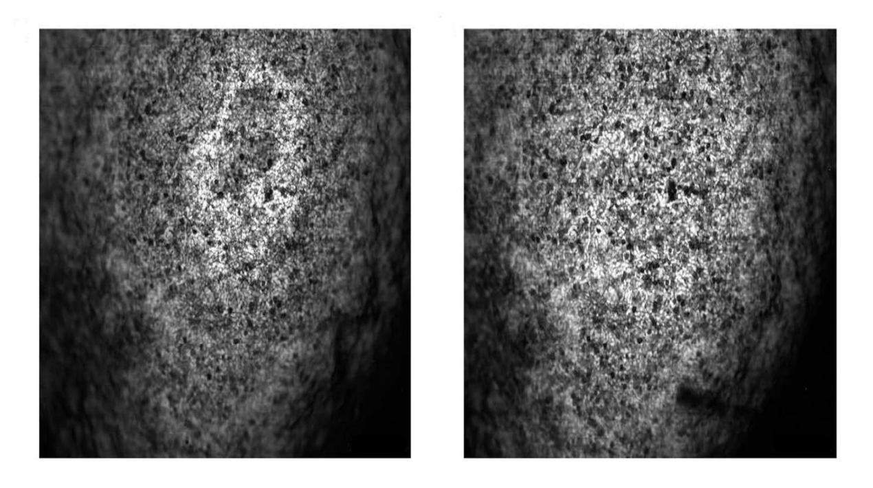
The researchers used two different imaging techniques to show this increased depth of field, including imaging in vivo mouse brain vasculature with a blue laser and imaging fresh organ samples with an ultraviolet laser.
According to Wang, this approach opens new possibilities for examining tissue samples during surgery, allowing total eradication of malignant cells and maximum preservation of normal ones. Translation into the operation room is a logical area for investigation in the future.
Meanwhile, the only core facility on the Caltech campus that offers light microscopy facilities is the Biological Imaging Facility or BIF (formerly known as the Biological Imaging Centre, BIC).
The BIF offers software tools for processing and interpreting image data in addition to microscopes. Two computer workstations at the BIF are equipped with Bitplane’s Imaris 3D/4D image processing and visualisation software.
A third computer workstation is running the Linux version of Scientific Volume Imaging B.V.’s (SVI) Huygens software, a strong image deconvolution application.


