


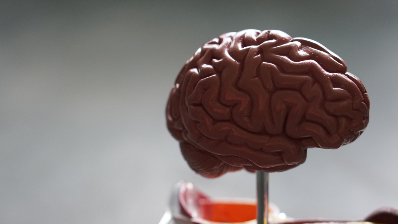
Scientists at the Indian Institute of Technology in Madras (IIT-Madras) and Massachusetts Institute of Technology (MIT) in the US, have grown human brain tissues called ‘organoids’ using a 3D Printed Bioreactor that they developed.
According to a report, the objective was to observe the brain tissues while they grow and develop, a technology that can potentially accelerate medical and therapeutic discoveries. The invention will help revolutionise treatment developments for diseases such as spinal cord injury, diabetes, rheumatoid arthritis, cerebral palsy, Alzheimer’s, Parkinson’s, and targeted cancer treatment. the organoid-based diagnosis is also useful for screening pharmaceutical compounds.
The current cell culture protocols involve separate chambers for incubation and imaging, requiring that cells be physically transferred to the imaging chamber, which poses the risk of false results and chances for contamination. However, the team has come up with a novel solution, which lets the cells grow uninterruptedly. A 3D-printed micro-incubator and imaging chamber was made into a single palm-sized platform, which was successfully demonstrated for long-term human brain cells culture and real-time imaging.
The scientists designed a microfluidic chip for the imaging and culturing of organoids. This ensures the long-term growth of organoids into a 3D spheroid form. A transparent glass disk with a thickness of 150m was placed on what researchers call a ‘Matrigel’ (extracellular environment matrix of polymers) that provided an optical window for live-organoid imaging. The chip was then put on a heating plate. The microfluidic chip was made using stereolithography-based 3D printing technology. It was refined using the 3D CAD modelling software. The printed chip was then cured by exposure to UV light and then connected to an incubator environment for the growth of organoids.
“The design from this research is a scalable microfluidic technology in which copies of an organoid can be grown simultaneously in different wells, for studies in basic and applied science,” Professor Anil Prabhakar, Department of Electrical Engineering, IIT-Madras, said in a statement. He added that the bioreactor can be completely automated with different protocols, and used for drug discovery, thus drastically reducing labour costs, errors, and time to market. Different environmental sensors can be combined with this micro-incubator and the device fits with most of the microscopes for live-cell imaging.
The technology has been patented in India. The research team is exploring the feasibility of international collaborations. The project was executed with support from the Centre for Computational Brain Research (CCBR) at IIT-Madras for funding and Sur’s Lab at MIT. Considering the importance of the micro-incubator in the field of healthcare and the pharmaceutical industry, the team is working through ISMO bio-photonics to develop a user-friendly minimum viable product and raising seed grants for its further development. This will enable biologists or laboratory technicians to operate, control, and monitor the growth of organoids with a user-friendly system powered by artificial intelligence (AI)-assisted automated cell culture protocol.
The findings of this research were recently published in the peer-reviewed international journal Biomicrofluidics. Cell culture is one of the fundamental steps in validation of the human organ model, whether it may be a pre-clinical study for COVID-19, cancer medicine discovery, or any other medicine for humans.


