


A technology is being developed at the University of Queensland (UQ) that will help address the shortage of trained pathologists in Australia.
According to a recent report, this rapid precision-scanning technology will be able to speed up medical diagnoses as well.
The work being done by the University’s Digital Pathology team will replace glass pathology slides with digital slides. Doing so would reap benefits such as faster analysis, distribution and storage.
According to the University’s researcher, a majority of GP diagnoses (70%) are based on pathology tests. The changes brought about by this new technology can revolutionise Australian laboratories.
The pace that Australia can train pathologists is much slower than the nation’s reliance and increased usage of the limited pathology services.
The demand has increased, therefore capacity should also increase. However, extending the work times should not be the solution.
Pathology will not be the only one to benefit from this breakthrough as the project could also have applications in immunology, histopathology and microbiology.
There is already a fully automated scanning system for immunology tests deployed and in use in a pathology laboratory.
Aside from this, the researchers are also working on new data management processes and delivery technologies.
This will allow the digitised slides to be stored indefinitely, thereby creating a potential data bank for further research and education.
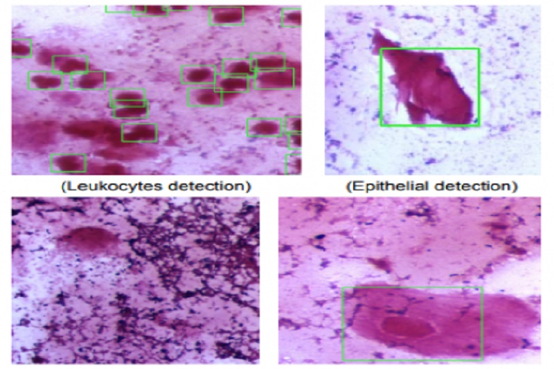
The team employs several technologies to create scanning and image-based Computer Aided Diagnostic systems.
These technologies include computer vision, machine-learning and pattern recognition methods.
Moreover, the group is being assisted by the University’s newest high-performance computer (HPC), Wiener, in their work. The University’s Research Computing Centre is also providing them support.
Wiener is equipped with the world’s most powerful graphics processing units (GPUs). These allow the team to train multiple machine-learning models using large data sets.
Wiener boosts the performance of the team as they are able to conduct the training five to eight times faster as compared to using the smaller legacy GPUs.
This allows the team to test several ideas at the same time.
The researchers create computer image data sets from different pathology areas and publish them after de-identifying patient data.
A startup, Viscient Pty Ltd, will continue the roll-out process and provide ongoing support of technologies developed from the project.
Wiener is designed to expedite the pace of research in a diverse range of imaging-intensive science, generated by the University’s world-leading microscopy facilities.
Wiener is a supercomputer that harnesses the capabilities of the most powerful GPUs ever made. It uses a mixture of deconvolution algorithms, machine learning and pattern recognition techniques.
Furthermore, it can provide near real-time outputs of de-convolved, tagged and appropriately characterised data.
Researchers can get immediate feedback on the quality of data being collected, allowing faster interpretation of microscopy data.


